
Introduction
The animal body is a complex structure made up of billions of cells. These cells are not randomly scattered but are organized into structural and functional units. In this chapter, we explore how cells form tissues, tissues group into organs, and organs coordinate to form systems, which together make up the body of an organism. Class 11 Ch2 Structural Organisation
Understanding this organization helps us appreciate how the body performs vital activities like movement, digestion, respiration, and reproduction in a coordinated manner. This chapter focuses on animal tissues, organ and organ system level of organisation, and the morphology and anatomy of selected animals – namely, earthworm, cockroach, and frog.
Class 11 Ch2 Structural Organisation
Levels of Structural Organisation
Living organisms exhibit different levels of organization:
- Cellular Level: Found in lower animals like sponges.
- Tissue Level: Cells performing similar functions form tissues (e.g., cnidarians).
- Organ Level: Different tissues combine to form organs (e.g., Platyhelminthes).
- Organ System Level: Groups of organs perform specific physiological functions (e.g., annelids, arthropods, chordates).
In higher animals, the organ system level of organization is most developed
3. Animal Tissues
Tissues are groups of cells with a common origin, structure, and function. In animals, tissues are classified into four major types:
a) Epithelial Tissue
These tissues cover body surfaces and line internal cavities.
- Types:
- Simple Epithelium: Single layer, found in alveoli, nephron, etc.
- Stratified Epithelium: Multiple layers, protective (e.g., skin).
- Subtypes of Simple Epithelium:
- Squamous: Thin, flat (e.g., lining of blood vessels)
- Cuboidal: Cube-shaped (e.g., ducts of glands)
- Columnar: Tall cells (e.g., intestine lining)
- Ciliated: With cilia to move substances (e.g., respiratory tract)
- Glandular Epithelium: Specialised to secrete substances like hormones, enzymes.
b) Connective Tissue
Connects and supports body parts.
- Types:
- Loose Connective Tissue:
- Areolar: Binds skin to muscles.
- Adipose: Stores fat.
- Dense Connective Tissue:
- Dense regular: Tendons (muscle to bone), ligaments (bone to bone).
- Specialised Connective Tissue:
- Cartilage: Flexible, in ear, nose.
- Bone: Rigid, supports body.
- Blood: Fluid tissue, transports substances.
- Loose Connective Tissue:
c) Muscular Tissue
Facilitates movement by contraction and relaxation.
- Types:
- Skeletal Muscle: Voluntary, striated.
- Smooth Muscle: Involuntary, non-striated (e.g., gut, blood vessels).
- Cardiac Muscle: Involuntary, striated, found in heart.
d) Nervous Tissue
Mainly composed of neurons and neuroglia, this tissue responds to stimuli and transmits signals.
- Neuron Structure: Dendrites, cell body (cyton), axon.
- Responsible for coordination, reflexes, and brain functions.
4. Organ and Organ Systems
In multicellular organisms, tissues form organs which combine into organ systems. Each system is specialized for a major function like respiration, circulation, digestion, etc.
- Example:
- Digestive System: Mouth, stomach, intestine.
- Nervous System: Brain, spinal cord, nerves.
- Reproductive System: Testes/ovaries and associated structures.
These systems work in coordination to maintain homeostasis.
5. Morphology and Anatomy of Selected Animals
The NCERT syllabus discusses the external and internal features of three animals as models: earthworm, cockroach, and frog.
5.1 Earthworm (Pheretima posthuma)
Morphology
- Shape: Long, cylindrical, segmented.
- Color: Brown with dorsal surface darker.
- Body Segmentation: ~100-120 segments.
- Clitellum: Thickened band from segments 14–16, important in reproduction.
- Setae: Bristle-like structures for movement.
Anatomy
- Digestive System: Straight tube; mouth → pharynx → oesophagus → gizzard → intestine → anus.
- Circulatory System: Closed type with blood vessels and hearts.
- Nervous System: Ventral nerve cord, ganglia.
- Reproductive System: Hermaphrodite (both male and female organs).
5.2 Cockroach (Periplaneta americana)
Morphology
- Color: Reddish-brown.
- Body Division: Head, thorax, abdomen.
- Wings: Two pairs.
- Antennae: Sense organs.
- Compound Eyes: For vision.
- Legs: Three pairs, jointed.
Anatomy
- Digestive System: Mouth → pharynx → crop → gizzard → intestine → rectum → anus.
- Circulatory System: Open type, with heart and sinuses.
- Respiratory System: Network of tracheae and spiracles.
- Nervous System: Brain, ventral nerve cord.
- Reproductive System:
- Male: Testes, seminal vesicles.
- Female: Ovaries, oviducts, spermatheca.
5.3 Frog (Rana tigrina)
Morphology
- Skin: Smooth, moist, without scales.
- Color: Dorsal side olive green, ventral side pale yellow.
- Body Division: Head and trunk (no neck or tail).
- Eyes: Bulging, with nictitating membrane.
Anatomy
- Digestive System: Mouth → buccal cavity → esophagus → stomach → small and large intestine → cloaca.
- Circulatory System: Closed type; three-chambered heart.
- Respiratory System: Skin, lungs, and buccal cavity.
- Nervous System: Brain, spinal cord, cranial and spinal nerves.
- Excretory System: Kidneys and urinary bladder.
- Reproductive System:
- Male: Testes and vasa efferentia.
- Female: Ovaries, oviducts.
Frogs are oviparous, with external fertilization and development through a tadpole larval stage.
6. Differences Between the Three Animals
| Feature | Earthworm | Cockroach | Frog |
|---|---|---|---|
| Body Division | Segmented | Head, thorax, abdomen | Head and trunk only |
| Circulation | Closed | Open | Closed |
| Respiration | Skin | Tracheae | Lungs, skin, buccal cavity |
| Reproduction | Hermaphrodite | Sexes separate | Sexes separate |
| Habitat | Moist soil | Dark corners, warm places | Freshwater |
7. Conclusion
Understanding structural organisation helps clarify how simple cells work together to form complex body systems in animals. This knowledge builds a strong base in biology, especially for future studies in anatomy, physiology, and zoology.
From the basic study of tissues to the internal structure of common animals, this chapter prepares students to understand life processes at an organismal level. Mastery of this chapter is not only essential for Class 11 exams but also critical for NEET and other competitive examinations.
Class 11 Ch2 Structural Organisation-NCERT TEXTBOOK QUESTIONS SOLVED
1. Answer in one word or one line.
(i) Give the common name of Periplaneta americana.
(ii) How many spermathecae are found in earthworm?
(iii) What is the position of ovaries in cockroach?
(iv) How many segments are present in the abdomen of cockroach?
(v) Where do you find Malpighian tubules?
Solution:
(i) Common name of Periplaneta americana: Cockroach
(ii) Number of spermathecae in earthworm: Four pairs
(iii) Position of ovaries in cockroach: Between 2nd and 6th abdominal segments
(iv) Number of abdominal segments in cockroach: Ten
(v) Location of Malpighian tubules: At the junction of midgut and hindgut in cockroach
2. What are the following and where do you find them in animal body?
(a) Chondrocytes
(b) Axons.
(c) Ciliated epithelium
Solution: (a) Chondrocytes – Chondrocytes are the only cells found in cartilage. They are present in spaces called lacunae and they produce and maintain the matrix of cartilage. Bending ability of cartilage is due to chondrocytes. Cartilage is present at tip of nose, pinna of ear, epiglottis etc.
(b) Axon – Axon is one of the processes of neuron, which is the structural and functional unit of nervous system. The part of cyton – n’here axon arises is axon hillock and axon ends in group of branches called terminal arborizations. It conducts impulses away from the cyton. Neurons (nerve cells)
are present in brain and spinal cord.
(c) Ciliated epithelium – If the columnar or cuboidal cells bear cilia on their free surface they are called ciliated epithelium. Their function is to move particles or mucus in a specific direction over the epithelium. They are mainly present in the inner surface of hollow organs like bronchioles and Fallopian tube.
More Resources for CBSE Class 11
- NCERT Solutions Class 11 Maths
- NCERT Solutions Class 11 Physics
- NCERT Solutions Class 11 Chemistry
- NCERT Solutions Class 11 Biology
3. Draw a labelled diagram of the reproductive organs of an earthworm.
Solution: 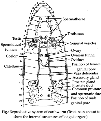
4. Answer the following.
(i) What is the function of nephridia?
(ii) How many types of nephridia are found in earthworm based on their location?
Solution: (i)
Nephridia are excretory organs in earthworms. Their primary function is to remove metabolic wastes, especially nitrogenous waste (like urea and ammonia), from the body. They also help in osmoregulation, maintaining the balance of water and salts in the earthworm’s body.
ii) There are three types of nephridia in earthworm based on their location:
- Septal Nephridia – Found on the septa between segments (from segment 15 onwards).
- Integumentary Nephridia – Located on the body wall throughout the body (more abundant on the anterior segments).
- Pharyngeal Nephridia – Located in the 4th, 5th, and 6th segments near the pharynx.
Each type plays a role in excretion and helps maintain internal fluid balance.
5. Draw a labelled diagram of alimentary canal of a cockroach.
Solution: 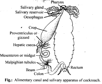
6. What are the cellular components of blood?
Solution: Blood is a fluid connective tissue. It is composed of plasma (fluid) and blood cells (corpuscles). Cellular components of blood (blood corpuscles) constitute about 45% of blood volume.
Three types of blood cells are:
(i) Erythrocytes or red blood cells: They are most abundant blood cells. Normal RBC count is 5-5.5 million/mm3 in males and 4.5-5 million/mm3 in females) RBCs help in transport of gases and maintain blood pH.
(ii) Leucocytes or white blood cells: The normal WBC count is 5000-6000/mm3 of blood. They are involved in immune response of body and act as soldiers and scavangers.
(iii) Thrombocytes or blood platelets: There are about 2,50,000 platelets/mm3 of blood. They are involved in blood clotting.
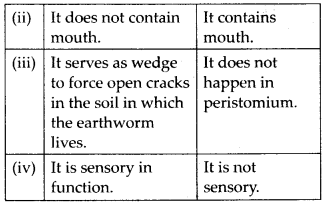
7. Distinguish between the following:
(a) Prostomium and peristomium
(b) Septal nephridium and pharyngeal
Solution: (a) Differences between prostomium and peristomium are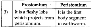
(b) Differences between septal and pharyngeal nephridia are: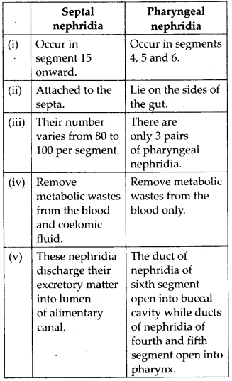
8. Mark the odd one in each series.
(a) Areolar tissue; blood; neuron; tendon
(b) RBC; WBC; platelets; cartilage
(c) Exocrine; endocrine; salivary gland; ligament
(d) Maxilla; mandible; labrum; antennae
(e) Protonema; mesothorax; metathorax; coxa.
Solution:
(a) Neuron: Areolar tissue, blood and tendon are connective tissues while neuron is a part a nervous tissue.
(b) Cartilage: RBC, WBC and platelets are parts of vascular connective tissue while cartilage is skeletal connective tissue.
(c) Ligament: Ligament is a connective tissue.
(d) Antennae: Maxilla, mandible and labrum are mouth parts of cockroach while antennae are sense organs.
(e) Protonema: Protonema is a filamentous juvenile stage in life cycle of Bryophytes, while mesothorax, metathorax and coxa are appendages of cockroach.
9. Match the terms in column I with those in column II.
| Column I | Column II |
| (a) Compound epithelium (b) Compound eye (c) Septal nephridia (d) Open circulatory system (e) Typhlosole (f) Osteocytes (g) Genitalia | (i) Alimentry canal (ii) Cockroach (iii) Skin (iv) Mosaic vision (v) Earthworm (vi) Phallomere (vii) Bone |
Solution: (a) – (iii), (b) – (iv), (c) – (v), (d) – (ii), (e) – (i), (f) – (vii), (g) – (vi)
10. Mention briefly about the circulatory system of earthworm.
Solution: Earthworm possesses a closed type of blood vascular system, as the blood flows through closed blood vessels. Blood is red in colour due to respiratory pigment haemoglobin. Prominent blood vessels in earthworm includes dorsal, ventral, sub- neural, lateral oesophageal and supra- oesophageal blood vessels. There are four pairs of tubular hearts, provided with valves. The anterior two pairs of hearts, known as lateral hearts lie in the 7th and 9th segments and connect the dorsal blood vessel with the ventral blood vessel. They receive blood from the dorsal blood vessel and convey it to the ventral blood vessel. The posterior two pairs of hearts are called latero-oesophageal hearts and are situated in the 12th and 13th segments. The latero-oesophageal hearts apart from connecting the dorsal and ventral blood vessels are also joined with the supra oesophageal blood vessel. Latero-oesophageal hearts carry blood from the dorsal vessel and the supra oesophageal vessel to the ventral blood vessel.Contractions keep blood circulating in one direction. Blood glands are present in the 4th, 5th and 6lh segments which produce blood cells and haemoglobin which is dissolved in blood plasma. Blood cells are phagocytic in nature.
11. Describe various types of epithelial tissues with the help of labelled diagrams.
Solution: Epithelial tissue is a tissue made of one or more layers of compactly arranged cells that covers external surface and internal free surface of body organs and which is underlined by a basement membrane. The various types of epithelial tissue along with the diagram are given below:
(i) Simple epithelium : It is composed of single layer of cells which rest on basement membrane. Simple epithelium generally occurs over secretory and absorptive surfaces and forms lining of body cavities, ducts and tubes. Simple epithelium is of several types.
(a) Squamous epithelium: It consists of single layer of flat cells, tightly linked together and have centrally located oval or spherical nucleus. It is also called pavement epithelium. It is found in walls of blood vessels, air sacs of lungs, and lining of eye lens.
(b) Cuboidal epithelium: Cells of cuboidal epithelium are as tall as wide, with centrally placed nucleus. Its main functions are secretion and absorption. It lines sweat gland, thyroid follicles, salivary glands. Brush bordered cuboidal epithelium, i.e., cells having microvilli on their free surface lines proximal part of uriniferous tubule, pancreatic duct, testis and ovary.
(c) Columnar epithelium: Cells are with basally located nucleus. It helps in secretion and absorption. It occurs in lining of intestine, stomach, gall bladder.
(d) Ciliated epithelium: Free surface of columnar and cuboidal cells are covered with cilia. Cilia help in moving fluids, particles, mucus, etc. in a specific direction. It occurs in the inner surface of Fallopian tubules, nasal passage, bronchioles.
(e) Pseudostratified epithelium: It consists of single layer of cells but some cells are shorter than others. Due to difference in size of cells, the epithelium appears 2-3 layered. Pseudostratified columnar epithelium occurs in urethra and parotid salivary gland. Pseudostratified columnar ciliated epithelium (only larger cells ciliated) occurs in lining layer of nasal’ chambers, trachea and large bronchi. It helps in moving mucus and foreign particles.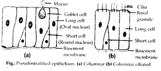
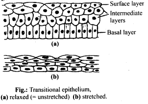
(ii) Compoundepithelium/stratifiedepithelium: It is multilayered epithelium where cells of only the lowermost or basal layer are in contact with basement membrane. It provides protection against mechanical and chemical stresses and has limited role in secretion and absorption. It covers dry surface of skin, moist surface of buccal cavity, pharynx, etc. Different types of compound epithelium are:
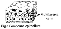
(b) Stratified cuboidal epithelium: The outer layer of cuboidal cells and basal layer of columnar cells. It lines ducts of sweat glands, large salivary and pancreatic ducts.
(c) Stratified columnar epithelium: Both upper and basal layers are made of columnar cells, e.g., epiglottis covering, part of urethra.
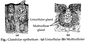
(iv) Glandular epithelium: It consists of specialised epithelial cells which synthesise intracellular macromolecules (protein in pancreas, lipids in adrenal glands, glycoprotein in salivary glands and all the three in mammary glands) and pour out the same in the form of a useful fluid secretion which is different from blood or any other extracellular fluid. Glands can be unicellular or multicellular on the basis of number of cells.
(a) Unicellular glands: Single-celled, e.g., goblet (mucous) cells of respiratory tract and alimentary canal.
(b) Multicellular glands: Consist of cluster of cells, e.g., Salivary glands.
On the basis of presence or absence of duct glands can be:
(a) Exocrine glands : These glands pour their secretion through a duct. They secrete milk, saliva, mucus, earwax. e.g., goblet cells, salivary glands, tear glands, gastric glands, intestinal glands.
Class 11 Ch2 Structural Organisation
Class 11 Ch2 Structural Organisation
Class 11 Ch2 Structural Organisation
Class 11 Ch2 Structural Organisation
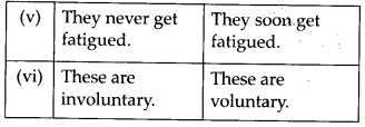
12. Distinguish between
(a) Simple epithelium and compound epithelium.
(b) Cardiac muscle and striated muscle.
(c) Dense regular and dense irregular connective tissues.
(d) Adipose and blood tissue.
(e) Simple gland and compound gland.
Solution:
(a) Differences between simple and compound epithelium are as follows: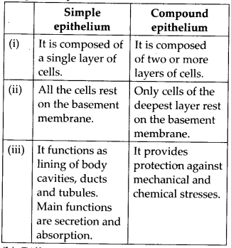
(b) Differences between cardiac and striated muscles are as follows: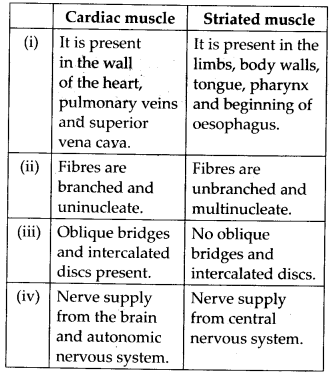
(c) Differences between dense regular and dense irregular connective tissues are as follows: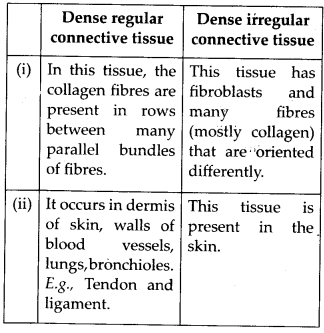
(d) Differences between adipose tissue and blood tissue are as follows: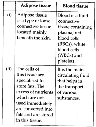
(e) Differences between simple gland and compound gland are as follows: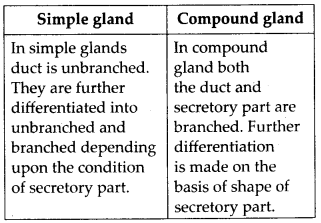
13. Draw a neat diagram of digestive system of frog.
Solution: 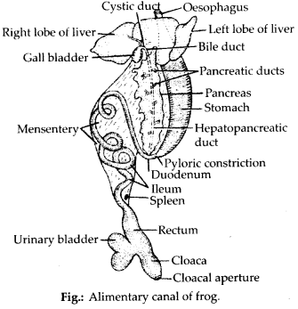
14. Mention the function of the following:
(a) Ureters in frog
(b) Malpighian tubules
(c) Body wall in earthworm.
Solution:
(a) Ureters in frog: Ureter is a transparent duct which arise from outer portion of kidney. In the “male frogs, ureter acts as urinogenital duct which runs backwards from kidneys and opens into the cloaca. It carries both urine and spermatozoa from kidney to the cloaca. In female, ureter conducts only urine from kidneys to the cloaca.
(b) Malpighian tubules: Malpighian tubules are excretory organs present in cockroach. These are present at junction of mid gut and hindgut. These are fine, long, unbranched, yellowish and blind tubules and are 100-150 in number. They help in the removal of excretory products from haemolymph.
(c) Body wall in earthworm: It consists of cuticle, epidermis, muscular layer and parietal peritoneum.
(i) It maintains the characteristic shape of’ the body.
(ii) It protects the internal organs.
(iii) The cuticle prevents excessive evaporation.
(iv) It serves as an ideal respiratory organ.
(v) The receptor cells play a vital sensory function.
(vi) The albumen helps in the formation of cocoon. It also serves as a food for the developing earthworm inside the cocoon.
(vii) Setae and muscles are responsible for locomotion.
(viii) Excretory matter is passed out through nephridiopores.
Class-Wise NCERT Solutions
Class 8
Class 9
Class 10
Oxford Science Solutions (Classes 6 to 8)
Periodic Assessment 1 (PA1) Class Test – Questions & Answers
Class 11 NCERT Solutions
- Class 11 Physics Solutions
- Class 11 Chemistry Solutions
- Class 11 Biology Solutions
- Class 11 Maths Solutions
Class 12 NCERT Solutions
- Class 12 Physics Solutions
- Class 12 Chemistry Solutions
- Class 12 Biology Solutions
- Class 12 Maths Solutions
Class 10 Sample Papers with Answers (2018–2025)
- 2018 Sample Paper with Answers
- 2019 Sample Paper with Answers
- 2020 Sample Paper with Answers
- 2021 Sample Paper with Answers
- 2022 Sample Paper with Answers
- 2023 Sample Paper with Answers
- 2024 Sample Paper with Answers
- 2025 Sample Paper with Answers
NEET Biology MCQs – Solved Previous Year Questions
NCERT Resource Hub
Math & Science Solutions by Class
Class 10
Class 9
Class 8
Class 7
Class 6
Class 12
Class 11
- Class 11 Math Solutions
- Class 11 Physics Solutions
- Class 11 Chemistry Solutions
- Class 11 Biology Solutions
Class-wise Solutions
Class 12:
Class 12 Physics – NCERT Solutions
Class 12 Chemistry – NCERT Solutions
Class 11:
- Class 11 Physics – NCERT Solutions
- Class 11 Chemistry – NCERT Solutions
- Class 11 Biology – NCERT Solutions
- Class 11 Math – NCERT Solutions
Class 10:
Class 9:
Class 8:
Class 7:
Class 6:
Subject-wise Solutions
Physics:
Chemistry:
Biology:
Math:
- Class 11 Math – NCERT Solutions
- Class 10 Math – NCERT Solutions
- Class 9 Math – NCERT Solutions
- Class 8 Math – NCERT Solutions
Science:
- Class 10 Science – NCERT Solutions
- Class 9 Science – NCERT Solutions
- Class 8 Science – Oxford Solutions
- Class 7 Science – Oxford Solutions
- Class 6 Science – Oxford Solutions